Multiscale Mechanics of Skeletal Tissues
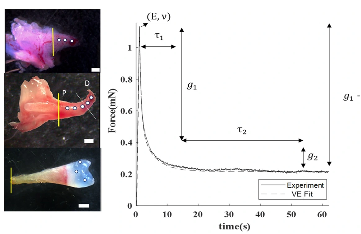
Mechanics of a regenerating axolotl limb
Axolotls regenerate their limbs. We are examining the mechanical properties of the tissue during regeneration using indentation and inverse finite element modeling. This will help us understand how the mechanical environment of the cells develops. Collaborator: James Monaghan
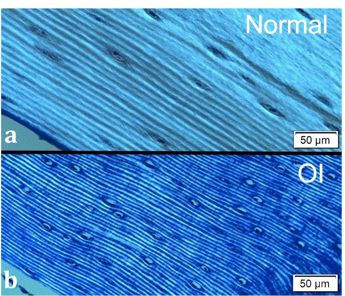
Osteon organization in osteogenesis imperfecta
Bone is organized in osteons (rings of bone surrounding a blood vessel). In the brittle bone disease osteogenesis imperfecta, which is caused by a genetic defect in collagen, the rings are thinner. This may contribute to the brittleness of the bone. Collaborator: Frederic Shapiro
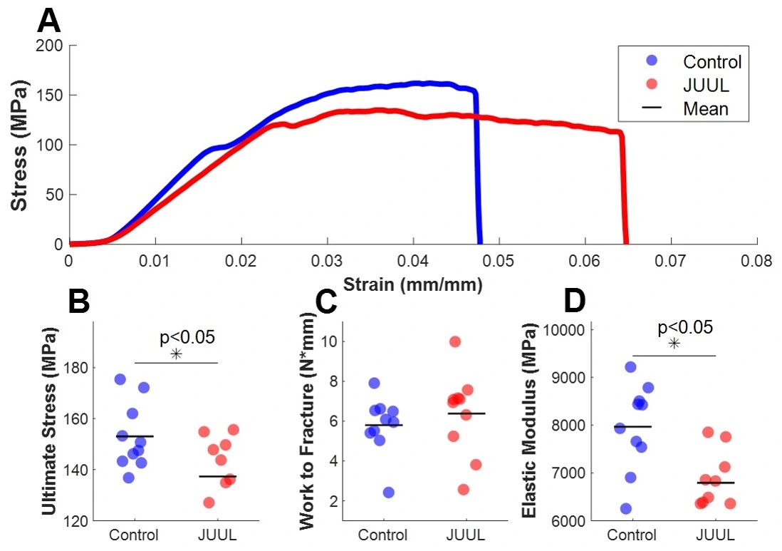
Vaping decreases bone toughness
We used a mouse of e-cigarrete exposure to determine the effects of vaping on bone. Electronic cigarettes alters bone structure and decreases the mechanical properties. Collaborators: Chiara Bellini and Jessica Oakes
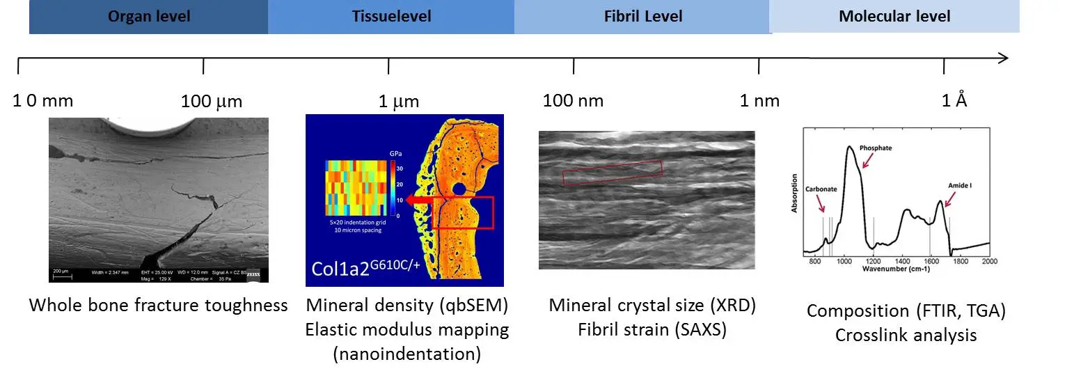
Multiscale toughness of bone
Toughness in bone arises from several mechanisms which act at multiple length scales in the bone hierarchical structure. In order to investigate the toughening mechanisms in bone, various parameters are measured in pathalogic mouse models of bone and related across the length-scales.
Previous Projects
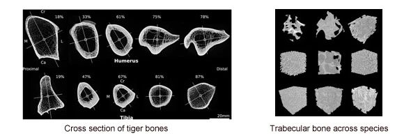
Bone scaling across species
Across animal species bone must support varied mass and locomotor activities. In this project we looked at bone scaling in the appendicular skeleton in felids (cats), artiodactyls (hoofed animals), macropods (kangaroos), and bipedal birds to understand how bone structure changes in response to increases in body size and locomotor habits. Collaborators: Dr. John Hutchinson (Royal Veterinary College)
Doube, M., Felder, A. A., Chua, M. Y., Lodhia, K., Kłosowski, M. M., Hutchinson, J. R., & S. J. Shefelbine** (2018). Limb bone scaling in hopping macropods and quadrupedal artiodactyls. Royal Society Open Science, 5(10), 180152.
M. Doube, S.C.W. Yen, M.M. Klosowski, A.A. Farke, J.R. Hutchinson, S. J. Shefelbine**, (2012). Whole-bone scaling of the avian pelvic limb. J Anatomy 221(1): 21-9.
M. Doube, M Kłosowski, A. M. Wiktorowicz-Conroy, J. R. Hutchinson, S. J. Shefelbine, (2011). Trabecular bone scales allometrically in mammals and birds. Proceedings of the Royal Society Biology B 278:3067-73.
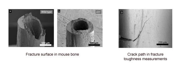
Characterizing fracture toughness in murine bones
Assessing bone quality is critical in understanding pathology and determining efficacy of therapy. Fracture toughness is a measure of how easy it is to break a bone. In this project we developed methods for creating resistance curves and calculating fracture toughness in mouse bones. Collaborators: Professor Rob Ritchie (U.C. Berkeley)
A. Carriero, E.A. Zimmerann, S. J. Shefelbine, & R.O. Ritchie, (2014). A methodology for the investigation of toughness and crack propagation in a mouse bone. Jouranl of the Mechanical Behavior of Biomedical Physics 39: 38-47.
A. Carriero, E.A. Zimmermann, A. Paluszny, S.Y. Tang, H. Bale, B. Busse, T. Alliston, G. Kazakia, R.O. Ritchie, S. J. Shefelbine, (2014). How tough is Brittle Bone? Investigating Osteogenesis Imperfecta in Mouse Bone. J Bone Miner Res 29(6): 1392–1401.